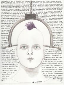
Once in the OR, they discussed the issues for getting me under (including awake intubation) and the final surgical plan...which was to include botox around the surgical site to lower how much it would spasm. It looked straight forward on paper, so I signed off and we were off to the intubation. If you want to read about the intubation, click the link...it is graphic so I am not assuming that people want to read it.
The last thing I remember after that whole mess is them saying that they had confirmed it was located correctly.
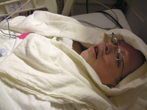
 Woke up in post op, relieved to have only a faint memory of them taking the intubation out and then another faint memory of them doing some quick scrubbing to get the blood off my face before Eliot came in. I could breathe fine. The anesthetic combo this time worked! What a relief considering the long intubation from the last surgery. And I wasn't too cold either.
It took forever to get a room...so I spent the better part of a day in recovery. When they finally got a room for me it was a double. The fragrance issues were going to be complicated.
The neurosurgeons barely spoke with Eliot post surgery as it went long and it was almost 8-8.30 pm when they told him they were done. Dr B swung thru the surgical waiting room on his way out & said it was a bad tethered cord. 8 on a scale of 1-10 and that someone would come to talk to Eliot. No one ever did.
I have the surgical report now. The surgery was based on the diagnosis of: Tethered Cord Syndrome in association with Noonan's syndrome, Ehler's-Danlos Syndrome (Beighton Score 8 with spontaneous dislocations of both hips and both shoulders) Chiari I Malformation with minimal tonsillar herniation; status post combined posterior fossa decompression and craniocervical fusion in extraction from the occiput to C4. They also list there being a lot of vascularized (arachnoidal) adhesions and that it was consistent with arachnoiditis.
They started out by puting me face down with bolsters under my shoulders & hips (no head support, which I certainly felt afterwards...I'm lobbying them to find a way to support people's heads!)...cleaning all around where they would make the roughly 6 inch incision. Not sure why, but they used iodine which I am allergic to... so I have a lot of blisters, rash & hives, still waiting to hear from them why they used the iodine. They did an xray to make sure of the right spot and marked it on my skin with indelible ink.
They performed the spinal cord untethering under color Doppler ultrasonography and fluoroscopic guidance (lots of good guidance tools to see what they needed to). And to get in to the area they needed to fix they did a laminectomy of L4 and a laminotomy of L3 & L5. Then they mapped everything...the arachnoid adhesions to cauda equina roots, and they were taut & packed into 2 tight lateral bundles. There were no fishtail movements and there was no measurable cerebrospinal fluid flow. They opened up the dura (basically a closed bag that surrounds the spinal cord & brain)...& so all the cerebrospinal fluid drains out (which is why I had to lay flat for a day to allow the CSF to regenerate after surgery and they put a lot of IV liquids in me later to assist with the regeneration). They removed all of the vascularized (abnormal or excessive formation of blood vessels) adhesions (the abnormal union of separate tissue surfaces by new fibrous tissue resulting from an inflammatory process) that were binding the various roots of the cauda equina & other areas...and sent some specimens to pathology.
When they cut the filum terminale, as they put it, the cut ends retracted briskly above and below the limits of the dural opening...about 5.5cm. They closed everything up with sutures, a paraspinal muscle graft & created an extradural blood patch.
When they did the final imaging, it revealed the nerve roots of the cauda equina to be greatly relaxed and evenly spaced throughout the lumbar theca. Good fishtail movements of the cauda equina elements were noted. The cut ends of the filum terminale were separated by a distance of 5.5cm There was 2.5-3cm/sec cerebrospinal fluid flow in the dorsal and ventral subarachnoid spaces, and between individual roots of the cauda equina. Cerebrospinal fluid flow was now moving well.
Beyond the excruciating pain of getting through the first few days, there was an extra pain at the top of my incision. They finally did an MRI & found there to be a hematoma. They say it will re-absorb of its own accord.

1 comment:
Thank you for posting all the details of your detethering surgery. I have an appointment a TCI in October and may end up doing a similar procedure. It is really valuable to get your patient perspective so I can make an informed decision should I get the chance.
Post a Comment